Anatomy Students View Bypass Surgery
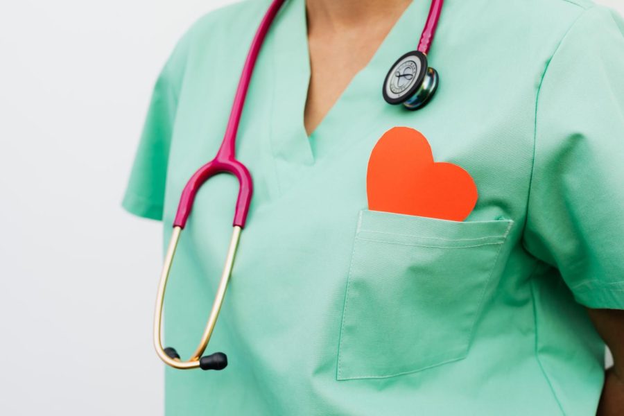
This year, anatomy students had the opportunity to enhance their learning of the cardiovascular system by watching a coronary bypass surgery. They gathered in the Innovation Center at LHS at the start of the school day on Friday, January 20, joined by anatomy teacher Jon Zaccaro. The presentation was live streamed via Zoom from the Liberty Science Center in New Jersey and was hosted by Director of STEM Innovation and Media Rosa Catala-Steidle.
The day started off with a brief overview of the surgery as well as different career paths in the medical field. Students learned that in addition to being a surgeon, one could become an emergency medical technician, sonographer, cardiovascular technologist, nuclear medicine technologist, and more. “[The presentation] helps introduce students to the long list of professions other than surgeon that are needed in an operating room,” said Zaccaro.
Then, Steidle listed the different staff members who are present in the operating room during a typical bypass surgery. The cardiothoracic surgeon performs the actual surgery and is assisted by a perfusionist, scrub nurses, and anesthesiologist. The perfusionist oversees the monitoring of blood flow and delivers medication into the bloodstream. The scrub nurses prepare the operating room and all of the instruments needed prior to the surgery, making sure nothing gets contaminated. They also aid the surgeon. Finally, anesthesiologists administer anesthesia—as well as an amnesic and paralytic—to the patient prior to the surgery. Zaccaro finds the role of the perfusionist especially interesting: “In this particular surgery, learning about the role of the perfusionist—who operates the heart/lung machine—was really interesting. The fact that this person is the reason the patient can stay alive without a beating heart is crazy to me.”
Steidle then reviewed the pathway of blood through the heart and delved into the specifics of a coronary bypass surgery. Officially known as a coronary artery bypass graft, the procedure is performed when there is a blockage of the blood flow to the heart. It involves taking healthy blood vessels from a separate part of the body—typically the chest, leg, arm, or abdomen—and connecting them to one of the coronary arteries, thus avoiding the blockage and restoring proper blood flow to the heart. “You take a vessel already in the human body and use it as a new pathway for the blood to flow,” Steidle explained. Other methods of treatment for a blocked artery are balloon angioplasty, stent implantation, and rotational atherectomy.
Then, the viewing of the surgery began. The procedure was pre-recorded at the Morristown Medical Center in New Jersey and was led by Head Surgeon John M. Brown. The patient, a 57-year-old female, suffered from hypertension and angina—or chest pain—and required four bypasses. After all of the medical staff scrubbed into the operating room, the patient’s skin was wrapped in plastic wrap to prevent pathogens from entering the wound.
The surgery began with an incision in the chest to provide access to the lungs and heart. The internal mammary arteries were harvested off of the chest wall. At the same time, an endoscope was inserted into the patient’s leg in order to gain a better view of the saphenous veins present there. Then, the veins were cut and sutured closed. The heart lung machine, which temporarily takes over the job of the patient’s heart and lungs by oxygenating the blood, was attached to the atrial appendage as the surgeon prepared to operate on the heart. Senior and anatomy student Alyssa Inserra found this part of the surgery especially interesting. “I never knew a thing like this existed, and it was fascinating to see how they could keep a person alive while her heart and lungs were stopped,” she said. The thoracic cavity was filled with water to cool it down, and after the water was suctioned out, the heart’s yellow color, the result of the fat surrounding its surface, was revealed.
The grafted veins and arteries were then sewn to the coronary arteries and heart in such a way that the blockages were avoided. Then, once blood was reintroduced to the heart and it started beating by itself, the heart lung machine was no longer needed. The chest was then sutured closed.
At the conclusion of the surgery, anatomy students had a chance to reflect on what they had seen and relate it to what they had been learning throughout the cardiovascular unit. “I always try to arrange the live surgery presentations during the current unit that it applies to. Not only does it give the students a real-life view of the organs and tissues that we are studying, but the disorder being treated—in this case, coronary artery disease—helps us understand the functions of that organ and how its damage can affect the life process it is supposed to support,” said Zaccaro.
Many students who take anatomy have an interest in pursuing a medical-oriented career in the future; thus, the surgery was a valuable tool and provided insight into what their jobs may entail. “This is really that one chance we can show students what they can do with the future that they envision, if that is in surgery/medicine. I hope it excites those that have that goal and even inspires some students who were unsure what the possibilities are,” said Zaccaro.
Senior and anatomy student Ethan Palacio hopes to be an anesthesiologist in the future and thought that the surgery was a “spectacular experience.” He added, “I loved being able to see the surgeon’s work in action.” Inserra also hopes to pursue a career in the medical field. “After taking anatomy for one semester of school, it really piqued my interest in the human body [and] its functions and capabilities,” she explained. “After watching the surgery, I knew that the medical field would be the place for me.”
However, that was not the case for all anatomy students. Junior and anatomy student Kerry Cullen, who does not have an interest in the medical field, found the surgery to be a little “too much,” but she was still able to understand how it could be helpful to others. “As a tool for learning about how surgery works, it was very useful and helpful,” she said. “Now, I know what everything looks like, rather than just hearing about it.”
All in all, the surgery allowed students to bring their learning about the cardiovascular system to life and expand their knowledge about the topic. “I always love to learn something new,” Zaccaro said. “I am consistently amazed by the work these doctors and nurses do, but when I learn about something I didn’t know before, it helps so much when I can relate the material we are discussing in a different way to my students.”
I am a member of the Class of 2023 as well as one of the editors-in-chief of the print edition of Horizon. I enjoy reading, playing the violin, and using...

